The subject of medical robots normally accompanies the assumption that they will soon replace clinicians. Those employed in healthcare know a different truth: healthcare embodies the repetitive and risk-heavy work for which robotics was developed. Anyone who has worked in a hospital understands the demand for patient care exceeds the amount of time you have in a day. This combined with ever-present staffing shortages and increasingly sick patients leads to physically demanding work which wears at the human body.

Enter the incredible innovations which have arrived with robotic-assisted surgery. These surgical systems are designed to reduce medical errors, assist surgeons with difficult fine motor tasks, and reduce the repetitive stress which occurs with prolonged surgeries.

A few of these incredible platforms are outlined below.

Da Vinci and Ion – Intuitive Surgical

Intuitive.jpg

source

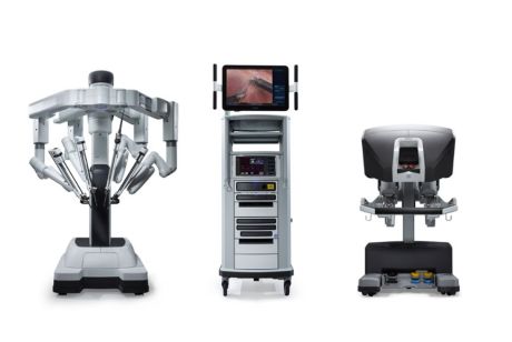
Intuitive Surgical was the first minimally invasive robotic surgery to come to market after being founded in 1995. The da Vinci surgical robot was first cleared by the FDA for general laproscopy, and through the years Intuitive has introduced several different da Vinci systems which allow surgical assistance for complicated procedures with precision and small incisions.

Instruments paired with the da Vinci system include clip appliers, suction and irrigation tools, needle drivers for suturing and closing incisions, and additional tools for dissection, coagulation, and cutting. This diversity in tasks allows a physician not only to perform tasks with precision and accuracy to ensure quality of a procedure, but also addresses healing time by reducing the size of incisions.

Additionally, the Ion system by Intuitive has been introduced and provides a catheter which is able to navigate the peripheral lung space for performing procedures such as biopsies.

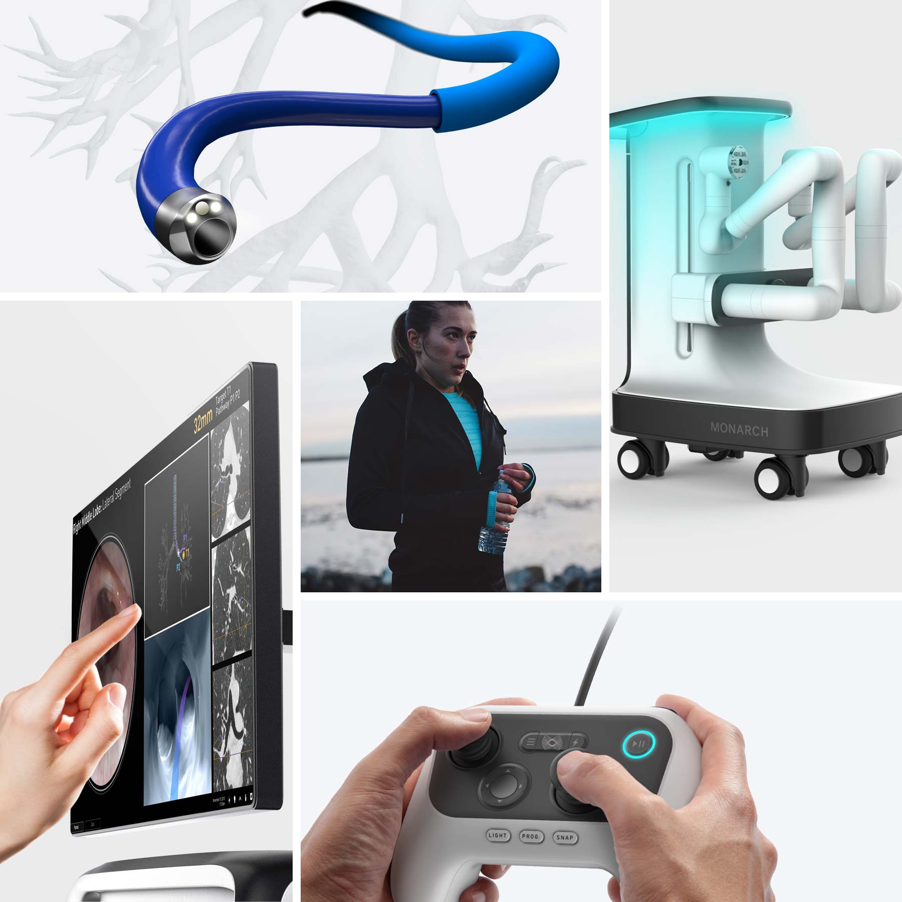
Monarch – Auris Health

home-robotics4-auris-health-collage-1800x1800.jpg

source

The Monarch platform provides a flexible, powered system for internal endoscopic procedures such as bronchoscopy. The current indication is to provide visualization and contact with patient airways for diagnosing and treating patients.

The platform is comprised of a robotic systems and its accessories, including a remote-power user interface which allows physicians to control the movement of the endoscope. Ultimately the goal of the platform is to tackle diseases such as lung cancer by providing more efficient and minimally invasive techniques for surgeons.

AquaBeam – PROCEPT BioRobotics

aquabeam-machine-1.png

source

Aquabeam is a robotic water ablation designed for the treatment of BPH (benign prostatic hyperplasia). Like the other systems, Aquabeam provides a combined visualization and treatment system to maximize efficiency and precision in patient care.


seismic

source

The evolution of powered wearables has been exciting to watch, evolving from clunky and with limited application to sleek and multipurpose. For those that have been following SRI‘s development of Superflex, there has been some wait to see the development of the soft wearable exosuit.

With the termination of Superflex and the acquisition of Lumo Bodytech by SRI comes the strength of the new SRI-sponsored company Seismic. Seismic is a new product which integrates technology from Superflex and movement-based algorithms from Lumo Bodytech to assist with human movement with a soft robotic exosuit.

For anyone along the aging population spectrum, powered wearable technology will provide independence and support to sustain daily activities and exercise. Thankfully due to progress in wearables, the appeal is growing to the mass market for application of common tasks.

The most appealing wearables are those that are easily integrated into a user’s life without many additional steps. Can a person integrate the wearable into their daily routine without thinking too much about activating or wearing the product? Passive integration is generally best.

The robotic design integrated into Seismic’s wearable gear facilitates easy enhancement of movement and activities. A person can simply slip on the gear, and activate the desired function. The gear complements the muscular activity which is the source of our movements.

For those following the evolution of wearables, Seismic provides a pathway to the integration of wearable robotic gear to improve everyday activity.  Movement and mobility mean independence, and for those that need a little extra assistance, this type of wearable will be a lifechanger.

After working for years in different healthcare settings, I learned to appreciate simple, good design of medical products, and have experienced the frustration of poor design.

Most of the time we use a medical product is for an important reason, often putting the function of the product before the design in importance. However, this model of function before design often leads to unforeseen problems from misuse.

One of the most important factors for whether a product will be adopted is whether the intended user can actually use the product, and how easily. Even if something is a major engineering accomplishment, if it is difficult for a patient or provider to use it will either be misused or not used at all. Misuse due to confusion and poor design lead to poor adoption.

Below are some frequently used medical products, categorized by quality of design.

Good Design: Pulse Oximeters

Image result for pulse oximeter

source

A pulse oximeter is a small medical device to measure the oxygen saturation in a person’s bloodstream. Typically in a hospital setting, you want oxygen saturation to be above 92%. Lower values mean a person is not getting enough oxygen, and this can result in fainting, falling, and other risks.

The reason that pulse oximeters have a good design is the ease of use. One simple inserts their finger into the device, which then reads the oxygen saturation using infrared light through the capillaries in the hand, and displays the reading on an easy to read screen.

There is little confusion about how to use this device, and little confusion about how to read the result.

Poor design: Most Wheelchairs

wc

Source

Having worked extensively with individuals using wheelchairs, there are very few which have been properly adjusted or fitted for people. Manual wheelchairs by design are difficult to adjust, both for patients and healthcare professionals, leading to frustration and improper fitting and use. In the rush of an injury such as sudden paralysis,  people are often improperly fitted and rushed into buying a wheelchair which does not suit them.

Manual wheelchairs have multiple parts, which can be difficult to adjust and store. Additionally, the weight of the product creates a burden on users, especially with repetitive use. Lightweight models made of carbon fiber have begun to address these issues, but the price remains high for many people to afford.

For the amount of years wheelchairs have existed in healthcare and been an option for those with limited mobility, there is still a long way to go before wheelchairs become an easily used product for short or long term use.  

Improving Design: Prosthetics

Image result for below knee prosthetic

Source

Prosthetics, particularly for below knee amputees, is one area of healthcare which has significantly improved in the last couple of decades. Companies have adopted modular designs for fit, comfort, design, and safety. Compared to the wooden, heavy prosthetics of 20 years ago, new prosthesis allow individuals to walk and run. 

The true beauty of improvement in prosthetics is being brought to surface by research labs such as Hugh Herr’s lab at MIT, where projects are breathing new life into  prosthetics with added function, mobility, and proprioception.


Image result for ergonomics

source

The term “ergonomics” normally has a similar effect on people as the words “healthy lifestyle:” guilt and unattainability. It conjures the image of someone sitting stick straight at a desk or lifting a box like a robot. We hear about it, know we should follow it, and ultimately dismiss it due to inconvenience.

One of the main lessons I learned from treating hundreds of people with work-related injuries is that unless there is a very traumatic injury, it is normally repetitive stress which wrecks our bodies. Our bodies are designed for a variety of functions which are often not physically accommodated in most jobs. Arms are designed for manipulation during a variety of daily tasks, not for constant daily typing. Our legs are designed to alternate loads during walking, and not for prolonged standing or repetitive squatting in place. Our torso muscles and spine developed to hold us upright during walking, and prolonged sitting has known deleterious effects1.

When I started doing ergonomic evaluations, I would normally sound off about 10 ideal postures and mechanics to which people would politely nod and then forget about.

In knowing the importance of correct body mechanics and trying to make ergonomics accessible, I found two simple rules can help people decrease the risk of potential strains and sprains:

Keep frequently used items close to your body

The further away from your body an object is, the less efficient your muscles are and the risk of strain increases. Keep frequently used items “bent elbow” distance from your body. 

Change positions frequently

Our muscles fatigue when trying to hold us in place for prolonged periods. One way to break up this fatigue and allow some recovery time is to alternate positions.

If you can’t keep close items which you’re working with, and you must perform repetitive tasks for your job, then enlisting assistance is a good idea. An exoskeleton which passively assists during repetitive squatting, repetitive overhead lifting, and repetitive bending helps during those tasks when our body is vulnerable to injury.


source

How is it really that a person can learn to move again after a debilitating injury? When a neurological injury such as a stroke or spinal cord injury occurs, people are left wondering how to function again. It can seem like the end of movement as they know it, a death in the way that a person has adapted to navigating their environment for years.

Depending on the type and severity of a neurological injury, the results of such an event can vary greatly. A brain injury such as a stroke disrupts the way that the brain serves as a command center for muscles, altering the way a person has learned to move and feel. A traumatic brain injury can completely alter movement, perception, and personality, depending on the area of the brain affected. Degenerative diseases such as Multiple Sclerosis slowly attack a person’s central nervous system, causing them to have a gradual decline in physical and sensory function.

In physical therapy, clients and their families are understandably scared and frustrated after such injuries. How does a person move forward with a life-changing disease or event? The hope lies in the power of adaptation, and the ability of the human neural circuitry to rewire and allow someone to learn to navigate the world in a new way. This is called neural plasticity, and it is a powerful survival tool which optimizes our resilience in life.

The nerves in our bodies communicate with biological electrical signals much like common electrical wires. When one nerve wire is faulty, our body finds a way to establish a signal through a new route so that a message can be delivered. This is absolutely amazing, and allows people to move forward in life after a debilitating disease or injury.

People often wonder how long it will take until they feel ‘normal’ again, and the beauty of neural plasticity is that our bodies find a new ‘normal’ through rewiring the neural circuitry. The key in this recovery is that the body and brain must be forced to find the new normal through practice and repetition. Encouraging someone to use their arm after a stroke, for example, fosters new signals in the body which recover upper body movement. Practicing walking after a stroke is vital in recovering walking ability.

The central nervous system recovers by creating new synapses. Where there was a blockage in communication from the injury, new receptors and new active signals are created. This requires increased stimulation in the brain, meaning that a person must be encouraged to do an activity which may seem difficult and new given their injury. It will initially feel like a new, uncoordinated task and will slowly become more efficient with practice as the brain and central nervous system adapt.

Peripheral nerves, the ones that transmit signals from the spinal cord to the rest of the body and back, also recover in several ways. The tail of the neuron, the axon, can regrow. In addition, a number of events are set off to create new signals and stimulate recovery.

Neuroprosthetics and technologies that force a person back into movement early may stimulate this mechanism of neural plasticity for an injured person. In general, the sooner that someone can start moving, the better. More time without movement means more muscle atrophy, as well as more of the body forgetting how to move through disuse.

The body responds to the commands and stresses that are placed on it, and devices and therapies that foster a plastic response are not only positive and productive, but what allow people to survive and adapt after a life-changing injury.

In some ways, a lasting traumatic brain injury (TBI) can be the worst kind of injury a person can endure in life. A TBI occurs when the brain is injured by force, and depending on the area affected, an individual is left with difficulty functioning and interacting with their environment. Our movement, sensation, communication, memory and learning is processed and controlled by the brain. When these functions are damaged, the interaction with our world is damaged as well.

Treating traumatic brain injuries may be one of the most emotionally and professionally difficult tasks. In an instant, the development of a person’s years of learning and communication can be erased from an injury such as a blow to the head, possibly leaving someone with the mental capacity and behavior of a toddler. Someone with a traumatic brain injury is often easily confused, unpredictable with their speech and actions, and occasionally aggressive as their frustration with communicating increases and appropriateness is inhibited. I recall treating an older gentleman who was the victim of violence, and just asking him to turn his head caused nausea, visual difficulty, and confusion in following just that simple command.

TBI’s can be divided into three main categories: mild, moderate, and severe. A mild injury often encompasses the kind of TBI that many people experience during their lifetime: a short-lasting concussion with possible loss of consciousness of up to a few minutes. Symptoms are often absent or mild, with some resulting nausea or headache. In a moderate injury, the force of the injury is greater and someone can be unconscious for up to 15 minutes with more lasting symptoms.

The third category is severe TBI’s, which can result from an event such as gunshot wound or the force of an explosion. Severe TBI’s cause permanent damage to the brain and leave lasting effects from which a person generally does not every fully recover. These chronic and lasting effects greatly affect a person’s ability to move, work, communicate with people, and function in society.

Until recently, treatment for people with chronic TBI’s was limited, but there is now hope for progress. A recent study by Chou et al introduced ISRIB, a drug tested in UCSF’s lab which was able to reverse the effects of a TBI in mice. This was done by inhibiting a stress response in the brain commonly associated with TBI. The integrated stress response has been shown to be chronically activated in someone with a TBI, affecting the hippocampus’ ability to store memory and influence healthy cognition. In addition, the drug was able to assist in synthesis of proteins which contribute to learning, in a process called Long Term Potentiation (LTP).

Because the effects of a severe traumatic brain injury can last for months or years before improving, results of treatment for TBI are often slow and inconsistent. Generally, there is no effective protocol treating chronic TBI’s because they are so varied in origin and presentation. This is why the breakthrough from UCSF is so impactive. To possibly reverse the effects of brain damage offers extensive hope and potential for TBI survivors, their families, and their care team.

The absolutely amazing aspect of ISRIB is that it affects chronic effects of TBI. Chronic effects of an injury are very difficult to reverse as the system has often acclimated to its chronic state, making the effects more stable and difficult to change. It is incredible that a drug has been developed to not only inhibit the pathway of a TBI, but reverse its deleterious and long-lasting effects. The potential implications of this medications are massive, possibly allowing people with TBI’s to not only restore memory but continue learning.

Thus far ISRIB has only been tested in mice, and the next phase is move it forward for human testing. ISRIB was licensed in 2015 to Calico, a California-based company which owns rights to discoveries in Dr. Walter’s biochemistry lab at UCSF.

BCI neurotech

source

How much effort goes into picking up a spoon? The planning and anticipation of which hand to use, where to place the hand, when to open and close the fingers, and how much weight to anticipate is complex and requires much coordination of the nervous and musculoskeletal systems.

In a normally functioning nervous system, movement of the extremities occurs when electrical impulses from the brain trigger a response which is sent to muscles. The central nervous system (brain and spinal cord) passes along electrical signals to the peripheral nervous system, and the nerves in the peripheral nervous system respond by communicating with their corresponding muscles.

When a person has a neurological injury causing paralysis, the signals between the central nervous system and peripheral nervous system are interrupted. Suddenly, simple every day tasks become complicated. An injury such as a fall causing quadriplegia can leave a person struggling to figure out how to move around and perform previously effortless everyday tasks such as eating and getting dressed. The aspiration with medical technology, then, is to make the transition from injury to adjustment as smooth as possible.

Neuroprosthetics are medical devices intended to assist with injuries to the nervous system. In recent years, there has been much growth with this technology using brain-computer interface (BCI), robotics, and exoskeleton technology. The challenge with neurological injuries, however, is that it is very difficult to replicate the intricate and precise workings of the brain and nervous system.

The team from BrainGate recently published a study following a quadriplegic subject in which they ultimately allowed him to use his brain to successfully control the movement in his arms to be able to feed himself. This amazing coordination of technology was achieved by implanting electrodes into his brain which picked up electrical signals and transfer these signals to Functional Electrical Stimulation (FES).

In this study, the electrodes implanted in the motor cortex picked up the electrical signals as he planned to use his upper extremities. The BrainGate system is able to decipher the signals from the brain activity and transfer it to the FES system through electrical pulses. These electrical pulses stimulated the muscles in his arm, creating the desired movement which the participant had planned for. Specifically, the man was able to feed himself using his hand for the first time in 8 years.

Still an investigational device, the BrainGate system is so promising in providing independence and versatility of movement, and the team is now working with the Harvard Wyss Center. The hope is that someday individuals will be able to implement neurotechnology such as this as soon as possible after injury, allowing for adjustment before the deleterious effects of immobility set in.

Watch the video below for more insight into this amazing work:


Displaying DSC_0031.JPG

Pneumonia is a potentially fatal infection of the lungs, causing them to accumulate fluid in the air sacs. Especially dangerous for the very young, old, and immunocompromised, it must be diagnosed and treated as quickly as possible. Currently, the gold standard for diagnosis is a chest x-ray, which is not only inconvenient and costly, but also exposes an individual to radiation.

A staple physician accessory has always been the stethoscope, a tool for amplifying sound when listening to the internal sounds of a patient. When a doctor is listening to your heart or lungs, this requires a combination of skill with placement and auditory detection to differentiate normal and abnormal sounds. This alone is not enough to diagnose a lung infection such as pneumonia, and thus a suspected diagnosis must be confirmed with an x-ray.

A new instrument looks to improve the accuracy and ease of diagnosing pneumonia while providing an inexpensive and convenient alternative to chest x-rays. Tabla works by streamlining a series of simple steps to detect possible lung infections. A provider places the device over a patient’s sternum, and then continues to move the stethoscope around known areas of the lung while a wireless app collects diagnostic data.

As medical instruments become digitized for accuracy, interpretation of patient data and output is becoming more standardized. Tabla is a brilliant device which not only streamlines the diagnostics process for lung infections, but eases the burden of cost and minimizes exposure to radiation in the treatment of pneumonia.

Related image

source

Those with spinal cord injuries (SCI’s) know that medicine still has a long way toward a successful solution for their injury.  Spinal cord injuries often occur as a result of trauma, such as a fall or gunshot wound. The initial physical compression and loss of blood supply to the spinal cord, followed by secondary edema and swelling cause a death of the spinal nerves which control our movement. In short, this type of injury usually takes away a person’s ability to walk and stand on their own.

SCI’s are normally classified in ASIA grades from complete (A) to normal (E), with incomplete injuries in between. Complete injuries involved complete loss of movement and sensation below the level of injury, while incomplete injuries maintain some preservation of sensation or motor control. Unfortunately, the rate of spontaneous recovery for those with complete injuries is low, while incomplete injuries have a slightly better success rate of recovery.

One project working toward a solution for spinal cord injuries by combining technology and rehabilitation is the Walk Again Project. Working toward a protocol for SCI recovery, this group has recently published research combining virtual reality and robotic assistance with variable gait training. And, it has shown promise of providing some recovery even for paralyzed individuals with complete SCI’s.

In the publication, the project demonstrates a partial return of neurological function in complete SCI’s by combining several methods of treatment. As the person controlled movement via a robotic exoskeleton with their brain using virtual reality for guidance, they also received some physical feedback from their environment. This physical feedback was applied to areas such as their feet or forearms in response to certain movements.

The results of this involved, year-long training are novel and incredible. People with previous complete loss of muscle and sensory function were able to regain some motor control, sensation, and proprioception after training. This is a novel publication by the length of the study and methods of guidance which lead those with SCI’s back toward recovery. The combination of brain machine interface, robotics, and rehabilitation provides a groundwork for future treatment options.

The effectiveness of this training may partly be explained by the idea that by forcing the body to walk and waking up the part of the brain which controls movement, the motor cortex, motor function is partially restored. Additionally, the physical movement may activate CPG’s (central pattern generators) in the spinal cord, which generate rhythmic movement. There may still be a long way to go toward medicine in SCI treatment, but this project provides solutions and hope through combined methods. Watch the video below for more insight into this amazing project:

source

One of the great challenges in biotechnology is interfacing synthetic materials with biological ones. Our bodies are designed with an extremely complex network of tissue, vascular, and neural structures to protect us and alarm us if there is something potentially dangerous to our system. If we sit for too long, for example, we feel discomfort and shift positions instinctively. If something is pressing against our leg and threatens to disrupt normal blood circulation, we perceive this threat with pain and pressure and respond accordingly.

Amputees and prosthetists have long been facing the issue of how to interface the residual limb with a prosthetic socket. Fitting for a prosthesis introduces a synthetic limb component to a biological one, and an improperly fitted socket can cause pain, pressure sores, and expose a residual limb to infection and tissue damage. And while there has been much improvement from the crude iron prosthetics that amputees once had to endure, there is still much room to improve to make the interface closer to a natural one.

One group at MIT has sought to address this disparity by developing a variable impedance prosthetic (VIPr) socket. Using MRI imaging and surface scanning techniques, researchers were able to find the tissue depth and where the socket was most likely to place pressure on the irregular bony areas of the residual limb. A socket was then 3D printed using this data to apply the least amount of pressure when fitted to the amputee.

After testing this socket on a below knee amputee, it was found that there was a 7-21% decrease in pressure on various bony areas of the leg compared to a regular socket during walking. While there is still no perfect socket or prosthetic interface for amputees, this is a step in the right direction to protect valuable and vulnerable human tissue.